A few years ago we conducted a field trial that led to a surprising finding of numerous cases of ulcerative enteritis and typhlitis (cecal inflammation) in preweaned dairy calves. Neonatal gastrointestinal (GI) disease is obviously a leading cause of high mortality in preweaned dairy calves, but the clinical manifestations associated with GI conditions tend to be less specific (watery diarrhea, inappetence, inanition, dehydration, reluctance to move, and fever) than the pathology documented in our study. A calf was considered to have died due to ‘ulcerative typhlitis’ (UT) if affected by grossly visible cecal ulceration (Figure 1) with or without perforation combined with a morphologic confirmation assessed through histopathology.
During a follow-up study on the same operation, an additional 103 calves were necropsied among which 49 were affected by UT. Age at death of calves was 11 days old on average although only 3 calves were ˃ 30 days of age (e.g., 33, 38, 54 days). Most of the deaths occurred during the first half of the second week of life. The typical cecal lesion was a focally extensive UT characterized by complete loss of the mucosa sometimes replaced by a thick coagulum of necrotic debris and fibrin, with partial loss of the submucosal layer and sometimes of the tunica. The deeper tissues were often affected by areas of coagulative necrosis, severe edema expanding the submucosa, and vasculitis and fibrinoid vascular necrosis with fibrin thrombi in small and medium caliber blood vessels. The transition from normal to lesioned tissue was often abrupt with a rim of moderate to abundant fibrinous inflammatory infiltrate comprised of degenerate and viable neutrophils and mixed to variably shaped bacteria.
PCR tests for bovine coronavirus, bovine rotavirus, and Cryptosporidium, and bacteriological cultures for S. enterica spp. were performed on fecal, cecum and ileum specimens from 96 of the examined animals (n = 43 cases with UT; n = 53 controls without UT). Among the identified infectious pathogens, bovine rotavirus was present in almost every calf and there was no difference (p-value = 0.12) in S. enterica prevalence between cases and controls. Interestingly, bovine coronavirus and Cryptosporidium actually occurred more commonly in controls as compared to animals with UT (p-value < 0.05) (Table 1).
Calf management on the operation included feeding twice a day with two liters of milk comprised of pasteurized waste milk and milk replacer blended to 13% solids, 22-24% fat, and approximately 28% protein. Medicated milk, mixed with neomycin and oxytetracycline (Neo-Oxy 100/100 MR, PharmGate, Omaha NE, USA), was fed therapeutically at the labeled dose (10mg/lb body weight/day) to all calves 5-12 days of age under veterinarian oversight. Starting at day 3 of age, a grain mix composed of pellets, molasses, and whole corn was added to the diet and gradually increased to 2.25 kg by the end of the first month.
Similar UT pathology has been documented previously in the literature (J Vet Diagn Invest. 2017 Mar;29(2):242-244) and described as cecal infarction (CI) with localized or diffuse peritonitis. Interestingly, in that study the authors stated that the Tulare branch of the California Animal Health and Food Safety Laboratory (CAHFS-Tulare), has diagnosed CI in calves ≤ 30 d of age for many years. Similar to our study, there were no differences in pathogens between cases and controls that would explain the disease process. Cecal infarction is a rare condition in human medicine and can be a result of occlusive or nonocclusive ischemic necrosis of antimesenteric portions of the cecum. However, in humans the lesion is usually encountered in aged, often debilitated individuals. The common factor may be the degree of physiologic impairment in both aged humans and calves.
It is possible that the severe inflammatory response observed in the cecum of these animals was the result of a disruption of the gut ecological balance. For example, dysbiosis within the gut environment has been indicated as having an important role in the pathogenesis of vasculitis because of the consequent intestinal permeability alteration and aberrant immune responses (Nature, 2016, volume 535, pages 65–74). Moreover, gastrointestinal vasculitis has been described in the context of primary systemic vasculitides (a rare group of inflammatory disorders, where damage is directed against the blood vessels), with intestinal infarction and perforation representing some of the main severe and life-threatening outcomes (Best Pract Res Clin Gastroenterol. 2005 Apr;19(2):215-33).
Unfortunately, the reality is that no one has been able to describe the “cause” of this particular pathology. I put cause in quotes because this gets at the heart of a concept we cover in 2nd year veterinary epidemiology. Causation comes down to necessary and sufficient factors. A necessary factor or factors MUST be present for a disease process. As an example, to the best of our knowledge Moraxella bovis is necessary for the development of infectious bovine keratoconjunctivitis (pinkeye). However, Moraxella bovis is not sufficient to cause disease. Other factors facilitate the disease process (e.g., damage to the cornea via grass awns, dust, etc.) such that the sufficient cause of pinkeye is multifactorial (e.g., Moraxella bovis + corneal damage). In fact, most sufficientcausal factors are multifactorial.
With that in mind, we really don’t know the necessary or sufficient factors that cause UT. That begs the question as to how close we have to be to the truth (the cause) to effect change. It turns out that (anecdotally) these UT lesions have been observed on at least two other operations that I know of. In those operations as well as the operation upon which we conducted our study there were consistent inconsistencies in the component parts of the milk mixture. Let me explain. As I pointed out above, the operation we worked with had a very consistent milk product in terms of solids, fat, and protein. And I can attest to the fact that they were also extremely consistent with the time of delivery, quantities fed, and temperature at delivery of the milk mixture. However, and this seems to be the crux of the situation, the various component parts that went into the milk mixture were inconsistent on a day-to-day basis in terms of the sourced milk (hospital versus bulk tank) and proportional amount of milk replacer required to standardize the mixture. This seems to have been the case as well with one of the other operations mentioned above. The third operation that I know of with similar UT issues was a small producer-processor and the UT arose during times when unsold milk was added back into the milk mixture which presumably changed the component parts in a similar fashion to the blended mixtures described above.
I’ll readily admit that there is anecdote built into this explanation but for the moment it is the closest thing to the cause of UT that I can describe. If in fact the sufficient cause of UT is an inconsistency in terms of milk mixtures, then we can potentially solve the problem without understanding exactly why the inflammation and pathology develop. Nonetheless, there is work in this area that may provide some useful insight into how the variations in composition between whole milk (WM) and milk replacer (MR) affect gastrointestinal development in young calves. For example, a recent paper (J Dairy Sci, Volume 106, Issue 4, p 2408-2427) compared the gastrointestinal tract structure and function in response to feeding WM or MR having the same macronutrient profile (e.g., fat, lactose, protein) to calves in the first month of life. Relatively little is known about how feeding WM versus MR affects the development of the GIT and intestinal permeability. In intestinal tissues, changes in permeability could be related to changes in mRNA expression of tight junction genes, and the fatty acid profile of the liquid feed has also been associated with changes in intestinal permeability and tissue composition in multiple species. Omega-3 (n-3) and omega-6 (n-6) fatty acids are thought to alter intestinal permeability due to their roles as anti- and pro-inflammatory precursors, respectively. Thus, it is plausible that composition differences between WM and MR mediate changes in gut permeability by facilitating changes in gene expression and tissue composition of GIT, which in turn alters intestinal permeability in calves. It is not too big a stretch then to envision inconsistent mixtures of WM and MR potentially leading to changes in the gut that ultimately manifest as UT. Time will tell what research in this area ultimately discovers, but in the meantime I’ll defer to the five C’s of calf management per Dr. McGuirk from the University of Wisconsin:
- Colostrum
- Cleanliness
- Comfort
- Calories
- Consistency
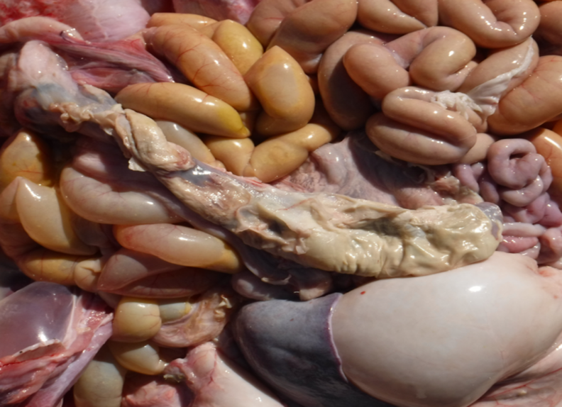
Figure 1. Ulcerative typhlitis in a two-week-old calf. Virtually the entire cecum is ulcerated and necrotic, with fibrinous peritonitis affecting abdominal organs.
Ulcerative Typhlitus
Table 1. Detection of intestinal pathogens from pre-weaned dairy calves affected by ulcerative typhlitis versus controls| Pathogen | Cases | Controls |
|---|---|---|
| Rotavirus | 43 (100%) | 49 (92%) |
| Coronavirus | 4 (9%) | 18 (34%) |
| Cryptosporidium | 1 (2%) | 12 (23%) |
| Cryptosporidium | 2 (5%) | 8 (15%) |
| TOTAL | 43 (100%) | 53 (100%) |