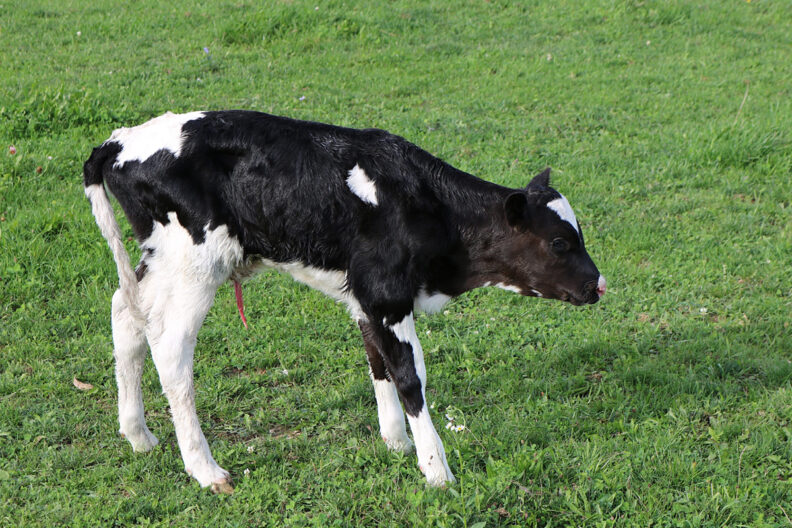
The umbilical cord may only be present for a few days after birth, but what happens during that short period can influence the health and productivity of a calf for weeks or even months. Omphalitis, commonly called navel ill, is an infection of the external umbilical stump and/or the internal umbilical structures (umbilical vein, arteries, and urachus). Although producers often think of omphalitis as an occasional problem, recent research suggests it may be considerably more common than previously appreciated.
Surprisingly, we currently do not know the true prevalence of omphalitis in U.S. dairy calves. The USDA National Animal Health Monitoring System (NAHMS) reports morbidity and mortality in preweaned calves but does not specifically track umbilical infections, making it impossible to estimate how common the disease is nationally. In contrast, studies from Europe have found omphalitis in approximately 20–31% of dairy calves (Meier et al., 2024), while studies in beef systems have reported prevalences exceeding 30% when calves are carefully examined using standardized clinical criteria (Perrot et al., 2024). This discrepancy likely reflects underdiagnosis rather than true biological differences.
Why does omphalitis matter?
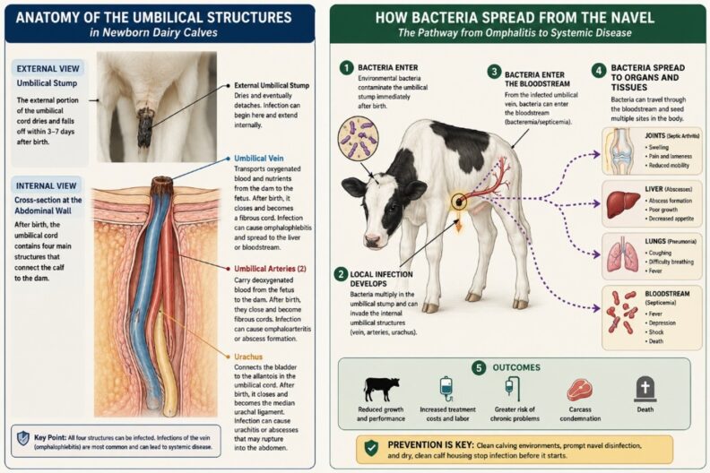
Omphalitis is more than a swollen or slow-healing navel. The umbilical stump can serve as an entry point for environmental bacteria during the first days of life. Infection may remain localized or extend internally through the umbilical vein, arteries, or urachus. In severe cases, bacteria can enter the bloodstream and spread to the liver, lungs, joints, and other tissues (Meier et al., 2024).
Affected calves may have pain, reduced milk intake, slower growth, and an increased risk of other diseases. Systemic spread can result in septic arthritis, pneumonia, liver abscesses, septicemia, and death. Omphalitis is also associated with greater antimicrobial use, treatment costs, and, in some cases, surgery or carcass losses (Perrot et al., 2024).
How can omphalitis be identified?
A normal umbilical stump should gradually dry, shrink, and detach. Calves should be examined for:
- swelling or abnormal thickening;
- heat or pain when the navel is gently palpated;
- moisture or purulent discharge;
- a firm, nonreducible mass;
- fever, depression, poor appetite, lameness, or swollen joints.
Visual examination alone may miss infections involving internal umbilical structures. Ultrasonography can help determine whether infection has extended along the umbilical vein toward the liver, the urachus toward the bladder, or the umbilical arteries (Wieland et al., 2017). There is currently no universally accepted diagnostic standard. Recent U.S. research suggests that measuring umbilical stump diameter with calipers may improve consistency when screening calves, although breed-specific thresholds and other clinical signs should also be considered (Rico et al., 2026). The latter study proposed two thresholds for stump diameter: one to estimate farm-level prevalence (16.5 mm for Holstein, 13.0 mm for Jersey) and another to identify calves likely to be diseased (19.3 mm for Holstein, 14.9 mm for Jersey).
Prevention
Prevention begins with reducing bacterial exposure immediately after birth. Calves should be born in a clean, dry calving area and moved to clean, dry housing. Wet or contaminated bedding increases exposure of the fresh umbilical stump to environmental bacteria (Perrot et al., 2024). A recent study of more than 3,400 dairy calves found omphalitis in 30.9% of calves and identified damp lying areas as an important risk factor (Meier et al., 2024). The navel should be completely dipped soon after birth using a farm protocol developed with the herd veterinarian.
Colostrum management remains essential for overall calf immunity, although recent studies have not consistently demonstrated that failure of passive transfer is an independent risk factor for omphalitis (Perrot et al., 2023).
A practical prevention program should include:
- clean and dry maternity pens;
- prompt removal of the calf from heavily contaminated areas;
- complete navel dipping rather than spraying only the outside;
- clean, dry calf bedding;
- routine navel checks during the first two weeks of life.
Treatment
Calves with an enlarged, painful, warm, or draining navel should be evaluated promptly. Early, localized infections are generally easier to treat than infections that have extended internally. Treatment should be directed by the herd veterinarian and may include systemic antimicrobials, anti-inflammatory medication, drainage of an abscess, or surgical removal of infected internal structures. Calves with fever, depression, lameness, swollen joints, respiratory signs, or poor appetite should be examined for systemic spread. Culture and antimicrobial susceptibility testing may be useful in severe, recurrent, or nonresponsive cases because several bacterial species can cause omphalitis and antimicrobial susceptibility can vary.
Take-home message
The national prevalence of omphalitis in U.S. dairy calves remains unknown because it has not been specifically measured in major national calf-health surveys. However, studies from other countries have reported omphalitis in approximately 20% to 30% of carefully examined calves, suggesting that the condition may be substantially underrecognized.
A clean calving environment, effective navel care, dry housing, and routine examination during the first weeks of life are the most practical steps for reducing its impact.