
By Drs. Chrissy Eckstrand and Christine Haake, Pathologists, WADDL
Gross Necropsy Description: A 9 kg, 3-week-old, intact male lamb was presented in fair postmortem condition and good body condition with moderate subcutaneous and visceral adipose tissue stores and moderate, bilaterally symmetric skeletal muscle mass.
Thorax: Pulmonary interlobular septa were mildly distended by clear fluid, and abundant white froth and mucus were present within the lumen of the trachea. Sections of lung floated in 10% neutral buffered formalin. There were approximately 20-25 mL of thin, watery fluid within the pericardial sac.
Urinary bladder: The bladder was moderately distended with urine. Approximately 500-1000 mg/DL glucose was detected in urine using a dipstick test.
Tissues also examined and considered free of significant gross lesions included the tongue, trachea, esophagus, thyroid, liver, spleen, kidneys, gastrointestinal tract, adrenal glands, skeletal muscle, bone marrow, eyes, testes, lymph nodes, and brain.
At this stage in the postmortem evaluation the primary gross diagnoses included 1) moderate, acute hydropericardium, and 2) mild, diffuse pulmonary edema along with moderate to marked glucosuria. Although the proximate cause of death was not evident on gross exam, the sudden death, age, and absence of significant gross abnormalities beyond pulmonary edema and hydropericardium suggested that enterotoxemia caused by Clostridium perfringens be considered as a possible cause of death. Furthermore, glucose detected within the urine via dipstick can be supportive of enterotoxemia. A small intestinal contents smear revealed numerous gram-positive rod bacteria (supportive of Clostridium).
Histopathology was conducted leading to the following histologic descriptions:
Brain: Virchow-Robins spaces surrounding blood vessels were expanded by increased clear space (perivascular edema). Multifocal, small, acute microhemorrhages were present throughout the grey and white matter of the midbrain, cerebrum, and cerebellum.
Lungs: Connective tissue surrounding pulmonary blood vessels was expanded by increased clear space (perivascular edema). Alveolar spaces were multifocally flooded with eosinophilic proteinaceous fluid (edema).
Small intestines: Moderately to markedly increased numbers of eosinophils were present multifocally within the lamina propria. In a separate section, the small intestinal lumen contained numerous degenerate neutrophils, mixed with ingesta and very high numbers of rod-shaped bacteria.
Tissues also examined and considered free of significant histologic lesions included the trachea, esophagus, heart, spleen, rumen, reticulum, adrenal glands, skeletal muscle, and bone marrow.
Based on this histologic findings the primary histologic diagnoses included 1) mild, acute, multifocal, perivascular edema in the brain and lungs, 2) mild, acute, multifocal pulmonary edema, 3) mild, peracute, multifocal hemorrhage in the brain, kidneys and thymus, and 4) moderate to marked, acute, multifocal, neutrophilic to eosinophilic enteritis within the small intestines.
The perivascular edema within the brain and lungs, multifocal small hemorrhages within the brain, kidneys, and thymus, and suppurative enteritis were supportive of enterotoxemia caused by Clostridium perfringens, which was grown in very large numbers in bacterial culture of intestinal contents. The causative agent of enterotoxemia, C. perfringens type D, was determined via toxin genotyping PCR as both alpha and epsilon toxin genes were detected. C. perfringens type D causes enterotoxemia in sheep (overeating disease). Predisposing factors include ingestion of excessive amounts of feed or milk in the very young and of grain in feedlot lambs, which allows overgrowth of epsilon toxin producing C. perfringens, causing systemic vascular damage. Acute death without clinical signs is common in lambs, though neurologic and respiratory signs may also be observed due to the presence of systemic edema. Given these findings it was suggested to the producer to review management, including vaccination of ewes prior to lambing, which is recommended for optimal prevention of this disease.
Necropsy investigation of acute death of a 3-week-old lamb
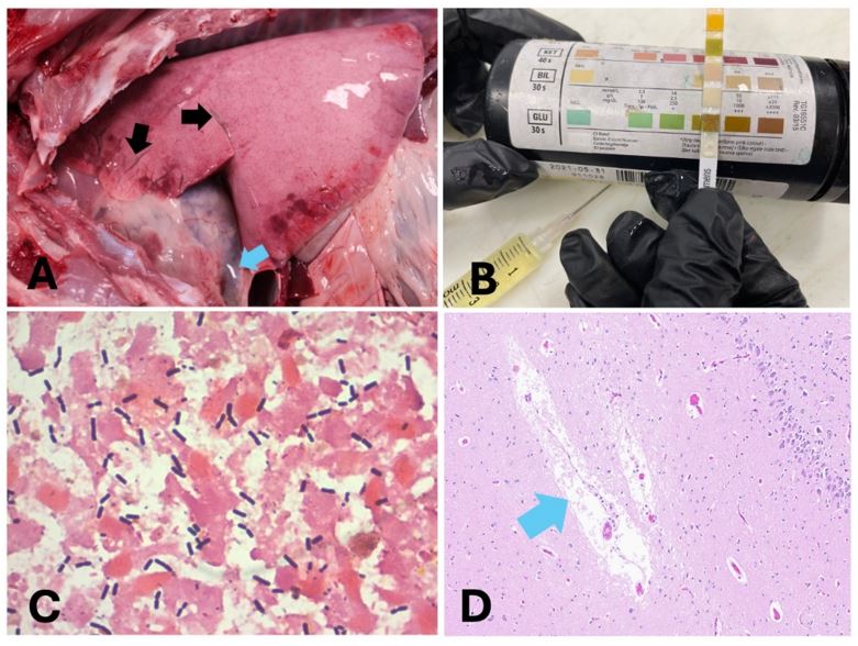
A. Necropsy revealed expansion of interlobular septa of the lung by clear fluid (pulmonary edema, black arrow) and increased pericardial fluid (blue arrow).
B. A urine dipstick revealed glucosuria.
C. Gram stain of an intestinal content smear revealed many gram-positive rod bacteria (supportive of Clostridium)
D. Histopathology of the brain demonstrates increased clear space around blood vessels (blue arrow, suggestive of cerebral edema)